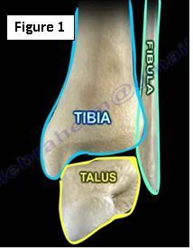
There are many of important topics associated with ankle fractures (Figure 1). One important topic is the classification of ankle fractures. There are two common classifications used for ankle fractures; the Weber Classification and the Lauge-Hansen Classification.

Weber Classification
The Weber Classification organizes the fractures of the ankle according to the level of the fibular fracture (Figure 2). Type A is a fracture that occurs below the level of the syndesmosis. This fracture is rarely unstable and is not normally associated with a syndesmotic injury. Type B is a common fracture that occurs at the level of the syndesmosis; this fracture could be unstable. Type C fracture occurs above the level of the syndesmosis and is usually unstable. A type C fracture with a deltoid ligament injury will most likely require syndesmotic screws because the syndesmosis will be unstable. The higher the fracture level, the more likely it will need syndesmotic screws.

Lauge–Hansen Classification
The Lauge-Hansen Classification depends on the mechanism of the injury. The mechanism is reliant on the position of the foot and the force that was applied to the foot (Figure 3). Along with supination and pronation, there is either an adduction, abduction or external rotation involved in the fracture mechanism of injury.

Supination-Adduction
A supination-adduction classification is described as a vertical medial malleolus fracture associated with an injury to the talus and tibial plafond. There is movement of the talus medially and a possible anteromedial tibial plafond impaction (Figure 4). There is also a transverse fracture of the distal fibula. Treatment includes a screw parallel to the ankle joint or an anti-gliding plate. The surgeon should check for loose bodies in the joint due to a possible tibial plafond impaction. They may also need to elevate and restore the joint surface. Fixation for this injury type may need to be started medially rather than laterally (routinely done laterally first).


Supination-External Rotation
The supination-external rotation is the most common injury. When looking at a fracture that needs to be classified, check the fibula on the lateral x-ray. This type of fracture will be noticed on the AP view and lateral view radiographs. On the lateral x-ray, if a fibular fracture that starts anterior/inferior and moves posterior/superior, this is an example of a supination-external rotation injury. This type of injury that can give you trouble if the fibula appears to be the only bone that is fractured because you want to prove that this is a supination-external rotation injury Type 2 and not a Type 4 injury. Make sure that the physician is not missing a Type 4 fracture of the medial malleolus or injury to the deltoid ligament. This injury has four stages: anterior tibiofibular ligament, fibular fracture, posterior tibiofibular ligament, and medial malleolus or deltoid ligament (Figure 5). Injury to the deltoid ligament may not show up clearly on x-rays. A stage 2 injury alone is treated with a boot and weight bearing as tolerated, while a stage 4 injury requires surgery.

Pronation-External Rotation
The pronation-external rotation is viewed on the lateral x-ray as a fracture that goes from anterior/superior to posterior/inferior. A fracture of the fibula is usually above the joint level (Weber Type C). This fracture has 4 stages starting medially. The fracture pattern moves in a circle similar to the supination-external rotation injury. The four stages being with medial malleolus or deltoid ligament, followed by the tibiofibular ligament, then the fibular above the syndesmosis, and lastly the posterior tibiofibular ligament (Figure 6).

Pronation-Abduction
A pronation-abduction injury is a fracture of the fibula that is usually transverse or comminuted (Figure 7). The fractured ankle may have injury only to the syndesmosis with nothing else appearing on the x-ray. This fracture will start medially and may cause injury to the deltoid ligament. Injury to the syndesmosis and fracture of the fibular will occur last.

For more information, visit my YouTube Channel:
Originally published at www.huffingtonpost.com