Introduction
There has been much furore regarding the gut microbiome, and many international academic and research centres have focused their research agendas on delineating the specific functionalities and therapeutic potentials of alterations in the gut microbiome. Innovative solutions are being pitched – from alterations to the composition of the gut microbiome, curated analysis of the gut microbiome to disease-targeting gut microbiome interventions; these solutions are predicated on the intricate relationship between the gut microbiome and the brain. In this article, I want to shed some light on the gut-brain-microbiome axis and dive deeper into this relationship.
The Gut Microbiome
The vast plethora of microbes which inhabit our gastrointestinal tracts, collectively termed the gut microbiome profoundly orchestrate and influence different facets of our physiology. An average-sized adult human body consists of approximately 10 trillion human cells. In stark comparison, the same body will harbour approximately 100 trillion microbial cells [19]. The gut microbiome contributes almost 2 kilograms of weight to the average sized adult human [10].
Furthermore, from a genetic standpoint, each of us contains about 20,000 genes, but as many as 2 – 20 million microbial genes. Each human body is a vast, rich, and complex ecosystem which is capable of influencing normal physiologic, immunologic, and metabolic processes. The human gut microbiome’s composition depends on a variety of factors including nutrition, mode of delivery, antibiotic usage, presence or absence of disease states (e.g., Celiac Disease, Inflammatory Bowel Disease, etc.) and physical activity amongst others.
For most humans, the maternal vaginal and perianal microbiota represent the first postnatal microbial exposure. A healthy human adult will harbour more than 1000 species of bacteria, amongst which the Bacteroidetes and Firmicutes phyla dominate [14]. Functionally, the gut microbiome is chiefly responsible for digestion and nutrition. For example, the capacity to digest xyloglucans which are found in certain vegetables was mapped to a single locus in a species of Bacteroides. This ability is a rare trait in Bacteroides – yet, 92% of human individuals harbour at least one of these rare Bacteroides species, conferring themselves with the ability to digest xyloglucans [14].
However, emerging research has shown that the gut microbiome is also capable of influencing the metabolism and immunology of the host. With respect to metabolism, several studies have shown that differences in energy consumption can be attributed to gut microbial composition. Individuals with a more efficient gut microbiome can extract dietary energy more efficiently and consequently may be predisposed to obesity [10]. Notwithstanding the gut microbiome’s physiologic and metabolic implications on the host, this paper’s primary focus will be on the potential health benefits of the gut-microbiota-brain axis on the central nervous system (CNS) and immune systems, particularly in chronic inflammatory processes.
Gut-Microbiota-Brain Axis
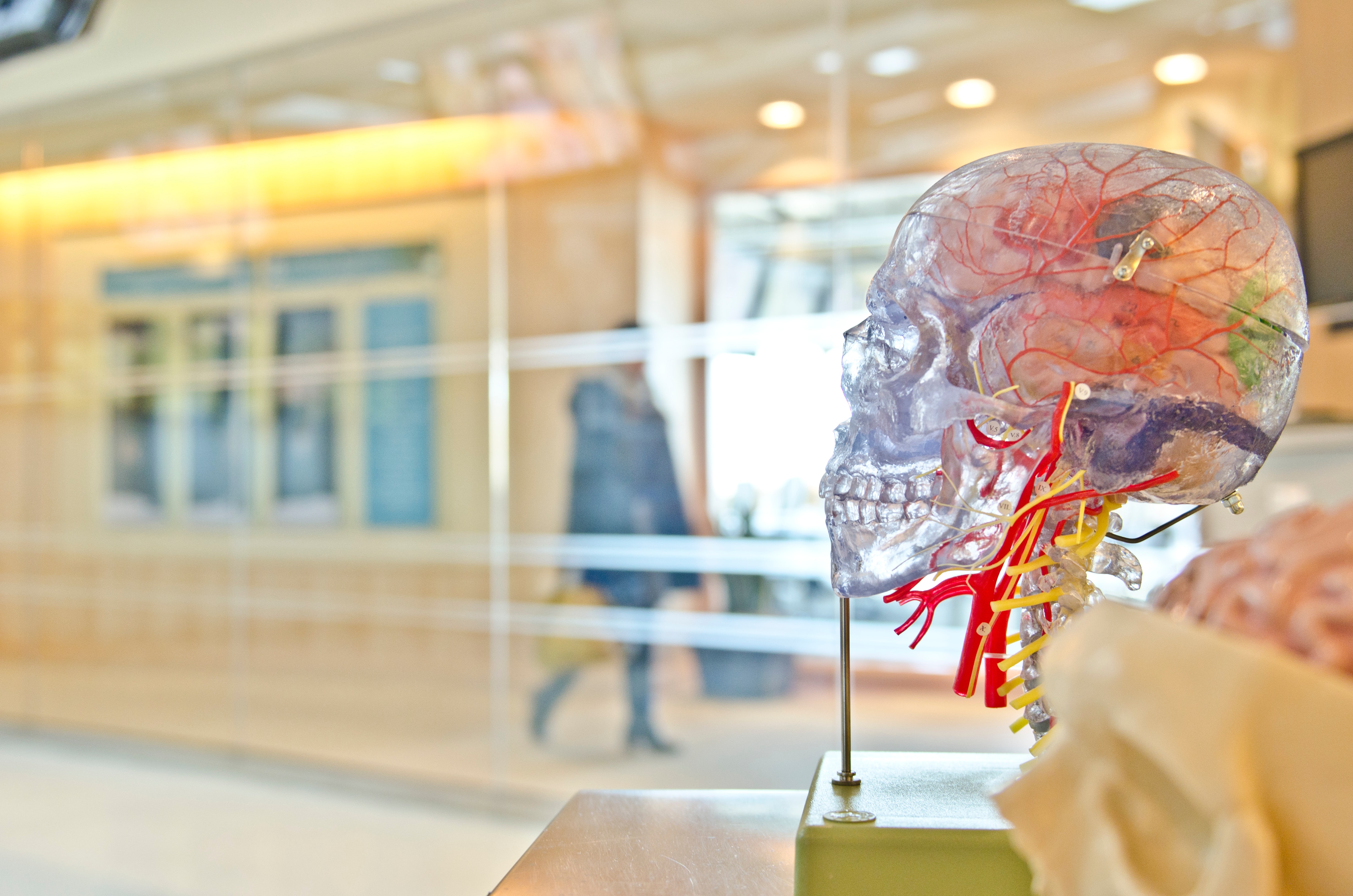
To define the gut-microbiota-brain axis, one must fully understand its individual components. The gut-microbiota-brain axis consists of the CNS, hypothalamic-pituitary-adrenal axis (HPA axis), the autonomic nervous system (ANS), the enteric nervous system (ENS), and the gut microbiome. The enteric nervous system (ENS), itself a division of the peripheral nervous system (PNS), is a complex entity comprising of myenteric, submucosal, and mucosal layers of neurons. Anatomically, it is large – comprising of more than 100 million neurons which form a complex network through the layers of the small and large intestines. Functionally, it is able to control gastrointestinal behaviour independently of the central nervous system. For example, the myenteric plexus which is localised between the longitudinal and circular layers of smooth muscle regulates smooth-muscle function, whereas the submucosal plexus which is localised in the connective tissue of the submucosa alters the secretory and absorptive capacities of the gastrointestinal tract. However, recent evidence shows that despite this autonomy accorded to the ENS by virtue of its intrinsic microcircuits, the gastrointestinal tract is still profoundly influenced by the CNS. This gut microbiome forms a symbiotic relationship with the brain by modulation of the CNS via several immune pathways. This relationship is bidirectional, in the sense that changes in CNS biochemistry can consequently alter the microbial composition via the HPA axis. This is achieved by altering the intestinal permeability to allow bacterial antigens to penetrate the epithelium, hence achieving an immune response in the intestinal mucosa [2]. Emotional and psychological stressors have also been shown to influence the composition of the gut microbiota, as evidenced by a study conducted in 2013 which found that healthy students exposed to stress had fewer Lactobacilli in their stool when compared to periods bereft of stress [4].
Dysbiosis and the CNS
Several studies have elucidated the potential benefits that the gut-microbiota-brain axis can confer upon the CNS. Probiotics such as VSL3, Bifidobacterium, and Lactobacillus induce Ly6C+ monocytes to increase neurogenesis and augment memory. They also exhibit anti-inflammatory properties and can attenuate mood disorders such as anxiety and depression [9]. A study conducted by Dinan, Stanton, and Cryan in 2013 also showed that the administration of “psychobiotics” – live microbes which confer health benefits when ingested by patients suffering from mood disorders. Another study conducted in 2002 observed a higher prevalence of Bifidobacteria in healthy children when compared to children with Autism Spectrum Disorder (ASD). However, children with ASD had a higher prevalence of Bacteroides Vulgatus and Desulfovibrio species in their stool [2].
The proposed mechanism for this observation is that dysbiosis (defined as a disruption in the normal homeostatic mechanisms which regulate the structure or function of the gut microbiota) leads to increased permeability and leakiness of the intestinal epithelium, hence allowing bacterial antigens and cytokines to enter the bloodstream and cross the blood-brain-barrier to affect behaviour [13]. A study conducted by Berer et al. in 2011 suggested that the gut microbiome may be implicated in the pathogenesis of Multiple Sclerosis (MS) – a chronic disease characterized by inflammation, demyelination, gliosis, and neuronal loss. Germ-free mice saw a delay in induced experimental autoimmune encephalitis (EAE), probably due to a mitigated inflammatory response. The amalgamated findings from multiple studies focusing on memory, mood, ASDs, and MS suggest that the gut-microbiome-brain axis represents a promising target for therapy in CNS pathologies.
Potential Benefits of the Gut-Microbiome-Brain Axis
To realise the reversal of dysbiosis, several strategies have been formulated to achieve the favourable state conferred by gut homeostasis. These treatment strategies include probiotics (live microorganisms), prebiotics (non-digestible fermentable oligosaccharides), dietary fibre (non-digestible carbohydrates of plant origin) and faecal microbiota transplantation (FMT). Probiotics are the most commonly used treatment strategy and have been shown to promote immune tolerance for certain allergies, specifically eczema [18]. However, probiotic intervention has not yet proved to be efficacious with regards to preventing other immune-mediated pathologies such as asthma [7].
Further research should be conducted on the timing of administration, as well as the specific constituents of the probiotic mixtures to achieve an optimal therapeutic index. A diet that is depleted of fibre has also been shown to be associated with a decrease in the Bacteroidetes levels in the gut [16]. Antibiotics have long been known to alter gut microbiota composition, and therefore are capable of inducing dysbiosis. FMT has been shown to be useful in treating antibiotic-resistant Clostridium Difficile induced diarrhoea [17]. but there is no concrete evidence that it is a viable treatment option for IBD despite modest results from several trials [15]. A controlled trial in the future may shed further light on this matter.
It is still not clear if the disparities in microbial variation seen in CNS pathologies and chronic inflammatory states are themselves a cause of the disease states or sequelae. What is clear is that reduced microbial diversity and specific variations in the microbiota are associated with these disease states. We also know that the gut microbiota is vital in exerting its physiologic, metabolic and immunologic functions and that dysbiosis can have remarkable ramifications for the host. Further research should be conducted to clearly establish the relationship between dysbiosis and disease states, as well as the therapeutic potentials of the treatment mentioned above strategies.
References:
- Berer, K., Mues, M., Koutrolos, M., Rasbi, Z., Boziki, M., Johner, C., Wekerle, H. and Krishnamoorthy, G. (2011). Commensal microbiota and myelin autoantigen cooperate to trigger autoimmune demyelination. Nature, 479(7374), pp.538-541.
- Carabotti, M., Scirocco, A., Maselli, M.A. & Severi, C. (2015). The gut-brain axis: Interactions between enteric microbiota, central and enteric nervous systems. Annals of Gastroenterology, 28(2), pp.203-209.
- de Goffau, M., Luopajarvi, K., Knip, M., Ilonen, J., Ruohtula, T., Harkonen, T., Orivuori, L., Hakala, S., Welling, G., Harmsen, H. and Vaarala, O. (2012). Fecal Microbiota Composition Differs Between Children With -Cell Autoimmunity and Those Without. Diabetes, 62(4), pp.1238-1244.
- Dinan, T. and Cryan, J. (2013). Melancholic microbes: a link between gut microbiota and depression?. Neurogastroenterology & Motility, 25(9), pp.713-719.
- Dinan, T., Stanton, C. and Cryan, J. (2013). Psychobiotics: A Novel Class of Psychotropic. Biological Psychiatry, 74(10), pp.720-726.
- Finegold, S., Molitoris, D., Song, Y., Liu, C., Vaisanen, M., Bolte, E., McTeague, M., Sandler, R., Wexler, H., Marlowe, E., Collins, M., Lawson, P., Summanen, P., Baysallar, M., Tomzynski, T., Read, E., Johnson, E., Rolfe, R., Nasir, P., Shah, H., Haake, D., Manning, P. and Kaul, A. (2002). Gastrointestinal Microflora Studies in Late‐Onset Autism. Clinical Infectious Diseases, 35(s1), pp.S6-S16.
- Fiocchi, A., Burks, W., Bahna, S., Bielory, L., Boyle, R., Cocco, R., Dreborg, S., Goodman, R., Kuitunen, M., Haahtela, T., Heine, R., Lack, G., Osborn, D., Sampson, H., Tannock, G. and Lee, B. (2012). Clinical Use of Probiotics in Pediatric Allergy (CUPPA). World Allergy Organization Journal, 5(11), pp.148-167.
- Frank, D., St. Amand, A., Feldman, R., Boedeker, E., Harpaz, N. and Pace, N. (2007). Molecular-phylogenetic characterization of microbial community imbalances in human inflammatory bowel diseases. Proceedings of the National Academy of Sciences, 104(34), pp.13780-13785.
- Fung, T., Olson, C. and Hsiao, E. (2017). Interactions between the microbiota, immune and nervous systems in health and disease. Nature Neuroscience, 20(2), pp.145-155.
- Mazidi, M., Rezaie, P., Kengne, A., Mobarhan, M. and Ferns, G. (2016). Gut microbiome and metabolic syndrome. Diabetes & Metabolic Syndrome: Clinical Research & Reviews, 10(2), pp.S150-S157.
- Merras-Salmio, L., Kolho, K., Pelkonen, A., Kuitunen, M., Mäkelä, M. and Savilahti, E. (2014). Markers of gut mucosal inflammation and cow’s milk specific immunoglobulins in non-IgE cow’s milk allergy. Clinical and Translational Allergy, 4(1), p.8.
- Murri, M., Leiva, I., Gomez-Zumaquero, J., Tinahones, F., Cardona, F., Soriguer, F. and Queipo-Ortuño, M. (2013). Gut microbiota in children with type 1 diabetes differs from that in healthy children: a case-control study. BMC Medicine, 11(1).
- Petra, A., Panagiotidou, S., Hatziagelaki, E., Stewart, J., Conti, P. and Theoharides, T. (2015). Gut-Microbiota-Brain Axis and Its Effect on Neuropsychiatric Disorders With Suspected Immune Dysregulation. Clinical Therapeutics, 37(5), pp.984-995.
- Shreiner, A., Kao, J. and Young, V. (2015). The gut microbiome in health and in disease. Current Opinion in Gastroenterology, 31(1), pp.69-75.
- Smits, L., Bouter, K., de Vos, W., Borody, T. and Nieuwdorp, M. (2013). Therapeutic Potential of Fecal Microbiota Transplantation. Gastroenterology, 145(5), pp.946-953.
- Thorburn, A., Macia, L. and Mackay, C. (2014). Diet, Metabolites, and “Western-Lifestyle” Inflammatory Diseases. Immunity, 40(6), pp.833-842.
- van Nood, E., Vrieze, A., Nieuwdorp, M., Fuentes, S., Zoetendal, E., de Vos, W., Visser, C., Kuijper, E., Bartelsman, J., Tijssen, J., Speelman, P., Dijkgraaf, M. and Keller, J. (2013). Duodenal Infusion of Donor Feces for Recurrent Clostridium difficile. New England Journal of Medicine, 368(5), pp.407-415.
- West, C. (2014). Gut microbiota and allergic disease. Current Opinion in Clinical Nutrition and Metabolic Care, 17(3), pp.261-266.
- West, C., Renz, H., Jenmalm, M., Kozyrskyj, A., Allen, K., Vuillermin, P., Prescott, S., MacKay, C., Salminen, S., Wong, G., Sinn, J., Stokholm, J., Bisgaard, H., Pawankar, R., Noakes, P., Kesper, D. and Tulic, M. (2015). The gut microbiota and inflammatory noncommunicable diseases: Associations and potentials for gut microbiota therapies. Journal of Allergy and Clinical Immunology, 135(1), pp.3-13.